Step into the next generation of cell biology with our intensive 3-day hands-on workshop on 3D Cell Culture, designed for students, researchers, and professionals seeking practical experience with modern in vitro models used in biotechnology, cancer research, drug development, and regenerative medicine.
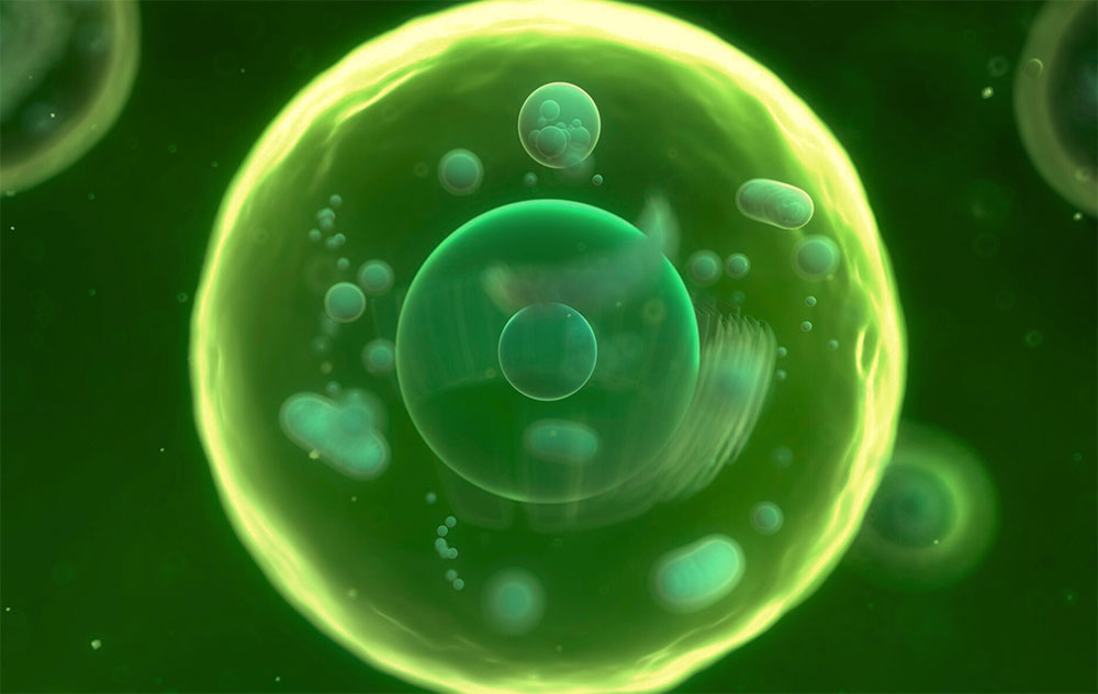
Traditional 2D cell culture systems often fail to accurately mimic the complexity of living tissues. In contrast, 3D cell culture models, including spheroids and microtissue systems, provide more physiologically relevant environments that better reproduce cell–cell interactions, tissue architecture, nutrient gradients, and drug responses observed in vivo. These advanced systems are increasingly becoming essential tools in translational research, toxicology, immunotherapy development, and precision medicine.
During this workshop, participants will gain both theoretical understanding and practical laboratory experience in the generation, handling, staining, imaging, and interpretation of 3D cell culture models. The course combines cell culture techniques, fluorescence microscopy, confocal imaging, live/dead staining, and experimental analysis in a highly interactive learning environment.
Participants will work directly with different 3D culture platforms, including hanging-drop systems, microcavity arrays, and specialized 96-well plates. Throughout the course, attendees will document and analyze their own experiments while learning how to troubleshoot common technical challenges associated with 3D cultures.
By the end of this workshop, participants will be able to:
This course is ideal for:
| Time | Activity | Format |
|---|---|---|
| 8:00 – 8:15 | Introduction & Course Overview | Lecture |
| 8:15 – 9:15 | Introduction to BSL-2 Guidelines | Lecture |
| 9:15 – 10:00 | Basics of 2D vs. 3D Cell Culture, Safety and Lab Organization | Lecture |
| 10:00 – 10:30 | Cell Culture Refresh: Harvesting, Counting and Adjusting Cell Density | Laboratory |
| 10:30 – 12:30 | Seeding 3D Cultures: Each participant seeds 1 Hanging-drop Plate (10 cm Petri Dish), 1 MicroSphere Array, and 1 Row of a faCellitate 96-Well Plate | Laboratory |
| 12:30 – 13:30 | Lunch Break | |
| 13:30 – 14:00 | History and Landscape of 3D Cell Culture: From Simple Aggregates to Organoids and Organ-on-Chip Systems | Lecture |
| 14:00 – 15:00 | Block 1 (Split Groups) • Group A: Trypsinisation and Cell Counting Practice on Individual 2D Culture Flasks • Group B: Introduction to the Fluorescence/Confocal Microscope (Controls, Focus, Filters, Best Practices) | Laboratory |
| 15:00 – 15:15 | Coffee Break | |
| 15:15 – 16:15 | Block 2 (Groups Switch) • Group B: Trypsinisation and Cell Counting Practice on Individual 2D Culture Flasks • Group A: Introduction to the Fluorescence/Confocal Microscope | Laboratory |
| 16:15 – 16:45 | Experiment Documentation and Outlook: Recording Seeding Parameters, Group Discussion, Q&A and Preparation for Day 2 | Discussion & Q&A |
| Time | Activity | Format |
|---|---|---|
| 9:00 – 9:20 | Welcome, Recap of Day 1, Lab Planning & Questions | Q&A |
| 9:20 – 10:00 | Basics of Fluorescence Microscopy | Lecture |
| 10:00 – 10:45 | Inspection of Participants’ Own 3D Cultures | Laboratory |
| 10:45 – 11:00 | Coffee Break | |
| 11:00 – 12:30 | Live/Dead Staining of Trainer-Prepared 72-Hour Spheroids: HepG2-GFP + PI + Hoechst or Alternative CellTracker Green + PI + Hoechst | Laboratory |
| 12:30 – 13:30 | Lunch Break | |
| 13:30 – 15:30 | Fluorescence Imaging (Depending on Group Size: Split Groups and Parallel Theory Sessions) | Laboratory |
| 15:30 – 16:30 | 3D Readouts and Assay Options; Group Discussion: Observations, Common Artefacts and Troubleshooting of 3D Cultures | Lecture & Discussion |
| Time | Activity | Format |
|---|---|---|
| 9:00 – 9:20 | Welcome & Open Questions | Q&A |
| 9:20 – 10:30 | Brightfield Imaging & Staining | Laboratory |
| 10:30 – 10:45 | Coffee Break | |
| 10:45 – 12:30 | Fluorescence Imaging (Depending on Duration and Sample Availability) | Laboratory |
| 12:30 – 13:30 | Lunch Break | |
| 13:30 – 14:30 | Preparation of Short Group Presentations (Optional Additional Fluorescence Imaging) | Theory |
| 14:30 – 15:30 | Group Presentations & Scientific Discussion | Theory |
| 15:30 – 16:00 | Final Wrap-Up, Feedback Session & Certificate Information | Q&A |
Duration: 3 Days
Training Type: Hands-On Laboratory Workshop
Location: Heidelberg, Germany
Class Size: Small groups to ensure intensive supervision and individualized support
Certificate: Participants receive an XTech Academy Certificate of Completion upon successful participation.
Reviews
There are no reviews yet.