Advance your expertise in 3D cell culture with this comprehensive hybrid workshop focusing on reproducible spheroid generation, real-time oxygen monitoring, hypoxia profiling, and functional mitochondrial analysis.
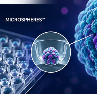
This intensive training program combines two online theory days with four days of hands-on laboratory training, providing participants with a complete workflow from spheroid generation to advanced functional characterization. Using MicroSpheres™, SensoSpheres™, MicrosCube™ oxygen sensing technology, fluorescence microscopy, confocal imaging, and mitochondrial stress testing, participants will gain practical experience with cutting-edge tools increasingly used in pharmaceutical research, cancer biology, toxicology, tissue engineering, and organ-on-chip development.
Unlike conventional 3D culture courses that focus primarily on morphology, this workshop emphasizes the functional biology of spheroids. Participants will learn how oxygen availability shapes cellular behavior, how mitochondrial function can be measured in three-dimensional tissues, and how physiological parameters can be integrated with imaging data to generate biologically meaningful insights.
Throughout the course, participants will design experiments, generate spheroids, perform staining procedures, collect real-time oxygen measurements, conduct mitochondrial stress tests, analyze their own datasets, and discuss how these technologies can be implemented in their own research projects.
This advanced course is recommended for participants who have:
By the end of this workshop participants will be able to:
| Time | Activity |
|---|---|
| 9:00 – 9:15 | Introduction & Course Overview |
| 9:15 – 9:45 | From 2D Monolayers to 3D Cell Culture |
| 9:45 – 10:30 | MicroSpheres: Principles & Experimental Design |
| 10:30 – 10:45 | Coffee Break |
| 10:45 – 11:15 | Staining Techniques in 3D: Live/Dead and Mitochondrial Probes |
| 11:15 – 11:45 | Microscopy & Imaging: Principles and Tips |
| 11:45 – 12:15 | LDH Assay as a Functional Readout in Spheroid Cultures |
| 12:15 – 12:30 | Q&A Session |
| Time | Activity |
|---|---|
| 9:00 – 9:15 | Welcome & Questions |
| 9:15 – 9:45 | Oxygen in 3D Cell Culture: From Hypoxia to Physioxia |
| 9:45 – 10:30 | Principles of Oxygen Sensing |
| 10:30 – 10:45 | Coffee Break |
| 10:45 – 11:15 | Experimental Design for Oxygen Measurement & 3D Mito-Stress Testing |
| 11:15 – 11:45 | Basic Data Interpretation |
| 11:45 – 12:15 | Overview of Practical Work & Hands-On Plan |
| 12:15 – 12:30 | Wrap-Up & Next Steps |
| Time | Activity |
|---|---|
| 8:00 – 8:30 | Welcome, Lab Planning & Questions |
| 8:30 – 9:30 | Introduction to BSL-2 Guidelines |
| 9:30 – 10:30 | Preparation of MicroSpheres & SensoSpheres; 2D Cell Harvesting and Single-Cell Suspension Preparation |
| 10:30 – 10:45 | Coffee Break |
| 10:45 – 12:30 | Completion of Cell Preparation and Parallel Seeding |
| 12:30 – 13:30 | Lunch Break |
| 13:30 – 14:15 | Calibration of MicrosCube Systems & Start of 72-Hour Oxygen Monitoring |
| 14:15 – 15:00 | Introduction to Fluorescence & Confocal Microscopy |
| 15:00 – 15:15 | Coffee Break |
| 15:15 – 16:00 | Open Lab & Questions |
| 16:00 – 16:30 | Daily Wrap-Up & Planning |
| Time | Activity |
|---|---|
| 9:00 – 9:15 | Short Recap |
| 9:15 – 9:30 | First Review of Oxygen Curves |
| 9:30 – 10:00 | Live/Dead Staining of Trainer-Prepared MicroSphere Spheroids |
| 10:00 – 10:15 | Coffee Break |
| 10:15 – 12:15 | Fluorescence & Confocal Imaging |
| 12:15 – 13:15 | Lunch Break |
| 13:15 – 14:15 | Brightfield Imaging of Participants’ Own MicroSpheres |
| 14:15 – 14:45 | Adjustment of Hypoxia/Hyperoxia Conditions |
| 14:45 – 15:00 | Coffee Break |
| 15:00 – 15:45 | Optional Imaging or Image Analysis Session |
| 15:45 – 16:15 | Q&A, Observations & Planning |
| Time | Activity |
|---|---|
| 9:00 – 9:15 | Recap & Planning |
| 9:15 – 10:00 | Imaging of Participants’ Own MicroSpheres |
| 10:00 – 11:15 | Hypoxia Staining: Fixation & Primary Antibody Incubation |
| 11:15 – 11:30 | Coffee Break |
| 11:30 – 12:30 | Optional Imaging or MitoTracker Staining |
| 12:30 – 13:30 | Lunch Break |
| 13:30 – 14:00 | Intermediate Oxygen Curve Analysis |
| 14:00 – 14:45 | Theory Session: 3D Mito-Stress Test |
| 14:45 – 15:00 | Coffee Break |
| 15:00 – 15:30 | Design of Mito-Stress Experiments |
| 15:30 – 16:15 | Future Outlook: Organoids & Organ-on-Chip Technologies |
| Time | Activity |
|---|---|
| 9:00 – 9:15 | Recap & Planning |
| 9:15 – 9:30 | Preparation of First Mito-Stress Test |
| 9:30 – 12:00 | Control 3D Mito-Stress Test |
| 9:45 – 11:45 | Parallel Hypoxia Staining: Secondary Antibody Incubation |
| 12:00 – 13:00 | Lunch Break |
| 13:00 – 13:30 | Preparation of Second Mito-Stress Test |
| 13:30 – 16:00 | Substance/Inhibitor-Based 3D Mito-Stress Test |
| 13:45 – 15:45 | Parallel Fluorescence & Confocal Imaging of Hypoxia-Stained Samples |
| 16:00 – 16:30 | Joint Discussion & Preliminary Data Review |
Format: Hybrid (2 Online Theory Days + 4 In-Person Laboratory Days)
Location: Heidelberg, Germany
Class Size: 4–6 Participants
Training Style: Intensive Hands-On Workshop
Certificate: XTech Academy Certificate of Completion
Recommended Background: Prior experience in mammalian cell culture and basic 3D spheroid culture systems.
Reviews
There are no reviews yet.